1.3 Meiosis
Stages of Meiosis
Most eukaryotes reproduce sexually — a cell from one individual joins with a cell from another to create offspring. To be successful, the cells that fuse must contain half the number of chromosomes as in the adult organism. Otherwise, the number of chromosomes would double with each generation, which would be unsustainable. The chromosome number is reduced through the process of meiosis. Meiosis is similar in many ways to mitosis, as the chromosomes are lined up along the metaphase plate and divided to the poles using microtubules. It also differs in many significant ways from mitosis.

Meiosis I and Meiosis II
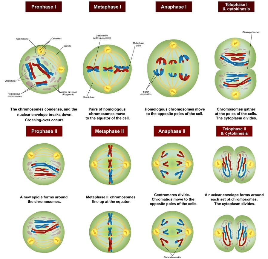
Meiosis has two main stages, designated by the roman numerals I and II. In Meiosis I homologous chromosomes segregate, while in Meiosis II sister chromatids segregate (Figure 1.3.2). Most multicellular organisms use meiosis to produce gametes, the cells that fuse to make offspring. Some single celled eukaryotes such as yeast also use meiosis to enter the haploid part of their life cycle. Cells that will undergo meiosis are called meiocytes and are diploid (2N) (Figure 1.3.3 and Figure 1.3.4). You will hear of cells that have not yet undergone meiosis to become egg or sperm cells called oocytes or spermatocytes respectively.
Meiosis begins similarly to mitosis in that a cell has grown large enough to divide and has replicated its chromosomes. However, Meiosis requires two rounds of division. In the first, known as Meiosis I, the replicated, homologous chromosomes segregate. During Meiosis II the sister chromatids segregate. Note how Meiosis I and II are both divided into prophase, metaphase, anaphase, and telophase, since those stages have similar features to mitosis. After two rounds of cytokinesis, four cells will be produced, each with a single copy of each chromosome in the set.
Meiosis I
Meiosis I is called a reductional division, because it reduces the number of chromosomes inherited in each of the daughter cells – the parent cell is 2N while the two daughter cells are each 1N. Meiosis I is further divided into Prophase I, Metaphase I, Anaphase I, and Telophase I, which are roughly similar to the corresponding stages of mitosis, except that in Prophase I and Metaphase I, homologous chromosomes pair up with each other, or synapse, and are called bivalents (Figure 1.3.5), in contrast with mitosis where the chromosomes line up individually during metaphase. This is an important difference between mitosis and meiosis, because it affects the segregation of alleles, and also allows for recombination to occur through crossing-over, which will be described later. During Anaphase I, one member of each pair of homologous chromosomes migrates to each daughter cell (1N) (Figure 1.3.4).
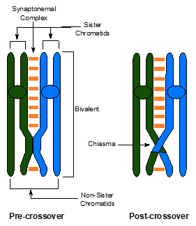

During Prophase I, the homologous chromosomes pair together and form a synaptonemal complex. Crossing over occurs within the synaptonemal complex. A crossover is a place where DNA repair enzymes break the DNA of two non-sister chromatids in similar locations and then covalently reattach non-sister chromatids together to create a crossover between non-sister chromatids. This reorganization of chromatids will persist for the remainder of meiosis and result in recombination of alleles in the gametes. Crossover events can be seen as Chiasmata on the synapsed chromosomes in late Meiosis I. Crossovers function to hold homologous chromosomes together during meiosis I so they orient correctly and segregate successfully. Crossing over also reshuffles the allele combinations along a chromosome resulting in genetic diversity, that can be selected in a population over time (evolution).
In Meiosis I, homologous chromosomes pair up, or synapse, during prophase I, line up in the middle of the cell during Metaphase I, and separate during Anaphase I. For this to happen, the homologous chromosomes need to be brought together while they condense during Prophase I. During synapsis, proteins bind to both homologous chromosomes along their entire length and form the synaptonemal complex (synapse means junction). These proteins hold the chromosomes in the transient structure of a bivalent (Figure 1.3.5). The proteins are released when the cell enters Anaphase I.
Summary of the Stages of Meiosis I
Meiosis I – A Reductional Division
Prophase I — Initially, chromosomes condense and become visible and centrosomes begin to migrate to opposite poles of the cell; Homologous chromosomes enter synapsis and the synaptonemal complex forms; Crossing over occurs resulting in an exchange of genetic material between non-sister chromatids of a homologous chromosome pair; Following this, the synaptonemal complex disappears and tetrads are visible; crossover points appear as chiasmata which hold non-sister chromatids together; finally, chromatids thicken and shorten, the nuclear membrane dissolves and spindle fibers begin forming.
Metaphase I — Tetrads line up on the equator or the metaphase plate and each chromosome of a homologous pair attaches to spindle fibers from opposite ends of the poles – sister chromatids attach to fibers from the same pole.
Anaphase I — Chiasmata dissolve; homologous chromosomes move to opposite poles; note: centromeres do not separate here.
Telophase I — The nuclear envelope reforms and the resulting cells have half the number of chromosomes, each consisting of two sister chromatids.
Interkinesis/Cytokinesis — Similar to interphase except no chromosome duplication occurs. Daughter nuclei become enclosed into separate daughter cells.
Meiosis II
At the completion of Meiosis I, there are two cells, each with one, replicated copy of each chromosome (1N). Because the number of chromosomes per cell has decreased (2->1), Meiosis I is called a reductional cell division. Meiosis II resembles mitosis, with one sister chromatid from each chromosome separating to produce two daughter cells. Because Meiosis II, like mitosis, results in the segregation of sister chromatids, Meiosis II is called an equational division (Figure 1.3.4).
Summary of the Stages of Meiosis II
Meiosis II – An Equational Division
Prophase II — Chromosomes condense, centrioles move towards the poles and the nuclear envelope disintegrates.
Metaphase II — Chromosomes align at the equator or the metaphase plate and sister chromatids attach to spindle fibres from opposite poles.
Anaphase II — Centromeres divide and sister chromatids move to opposite poles.
Telophase II — Chromosomes begin to uncoil; nuclear envelope and nucleoli begin to reform.
Cytokinesis — Division of the cytoplasm occurs, resulting in four new daughter cells, each containing haploid number of chromosomes.
Gamete Maturation
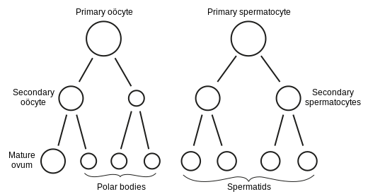
In animals and plants, the cells produced by meiosis need to mature before they become functional gametes. In male animals, the four products of meiosis are called spermatids. They grow structures, like tails, and become functional sperm cells. In female animals, the gametes are eggs. For each egg to contain the maximum amount of nutrients, typically only one of the four products of meiosis becomes an egg. The other three cells end up as tiny disposable cells called polar bodies (Figure 1.3.7). In plants, the products of meiosis reproduce a few times using mitosis as they develop into functional male or female gametes.
Media Attributions
- Figure 1.3.1 Meiosis main steps by Peter Coxhead, via Wikimedia Commons, CC0 1.0 Universal Public Domain Dedication
- Figure 1.3.2 Meiosis Stages by Ali Zifan, via Wikimedia Commons CC BY-SA 4.0
- Figure 1.3.3 Meiotic phenotype of blap75 mutants, Chelysheva et al. (2008), PLoS Genetics CC BY 4.0
- Figure 1.3.4 Original by Deyholos (2017), CC BY-NC 3.0, via Open Genetics
- Figure 1.3.5 Original by L. Canham (2017), CC BY-NC 3.0, via Open Genetics
- Figure 1.3.6 Chromosomal Crossover by Abbyprovenzano, CC BY-SA 3.0, via Wikimedia Commons
- Figure 1.3.7 Gray’s 7 (ovum maturation) by Fred the Oyster, public domain, via Wikimedia Commons
References
Canham, L. (2017). Figure 7. Diagram of a pair of homologous chromosomes during Prophase I [digital image]. In Locke, J., Harrington, M., Canham, L. and Min Ku Kang (Eds.), Open Genetics Lectures, Fall 2017 (Chapter 16, p. 6). Dataverse/ BCcampus. http://solr.bccampus.ca:8001/bcc/file/7a7b00f9-fb56-4c49-81a9-cfa3ad80e6d8/1/OpenGeneticsLectures_Fall2017.pdf
Chelysheva, L. et al. (2008) Figure 4. Meiotic phenotype of blap75 mutants [digital image], in The Arabidopsis BLAP75/Rmi1 Homologue plays crucial roles in meiotic double-strand break repair. PLoS Genetics, 4(12): e1000309. https://doi.org/10.1371/journal.pgen.1000309
Deyholos, M. (2017). Figure 6. Changes in DNA and Chromosome Content During the Cell Cycle [digital image]. In Locke, J., Harrington, M., Canham, L. and Min Ku Kang (Eds.), Open Genetics Lectures, Fall 2017 (Chapter 16, p. 5). Dataverse/ BCcampus. http://solr.bccampus.ca:8001/bcc/file/7a7b00f9-fb56-4c49-81a9-cfa3ad80e6d8/1/OpenGeneticsLectures_Fall2017.pdf
Fred the Oyster. (2020, October 30). File:Gray’s 7 (ovum maturation).svg [digital image]. Wikimedia Commons. https://commons.wikimedia.org/w/index.php?title=File:Gray%27s_7_(ovum_maturation).svg&oldid=507847250
Provenzano, A. (2013, 23 May). Chromosomal crossover [digital image]. Wikimedia Commons. https://commons.wikimedia.org/wiki/File:Chromosomal_Crossover.svg
Long Descriptions
- Figure 1.3.1 The stages of meiosis are numbered from 1 to 5. Cells initially contain a diploid number of 4 (2 grey and 2 pink indication homologous pairs). Step 1 shows DNA replication; step 2 shows pairing of homologous chromosomes; step 3 shows crossing over of non-sister chromatids; Step 4 shows production of 2 cells after Telophase 1; Step 5 shows production of four daughter cells with haploid number after Telophase 2. [Back to Figure 1.3.1]
- Figure 1.3.2 The various stages of meiosis in a cell containing 3 pairs of homologous chromosomes are as follows: Prophase I is shown as chromosomes condense, the nuclear envelope breaks down and crossing over occurs; Metaphase I is shown where pairs of homologous chromosomes line up at the equator if the cell; Anaphase I is shown where chromosomes migrate to opposite poles of the cell; Telophase I and cytokinesis is shown where cytoplasm divides; Prophase II is shown where spindle fibres form; Metaphase II is shown where chromosomes line up at the equator; Anaphase II is shown where centromeres divide and chromatids move to opposite poles of the cell, and finally Telophase II and cytokinesis is shown where nuclear envelope reforms and four daughter cells with a haploid number results. [Back to Figure 1.3.2]
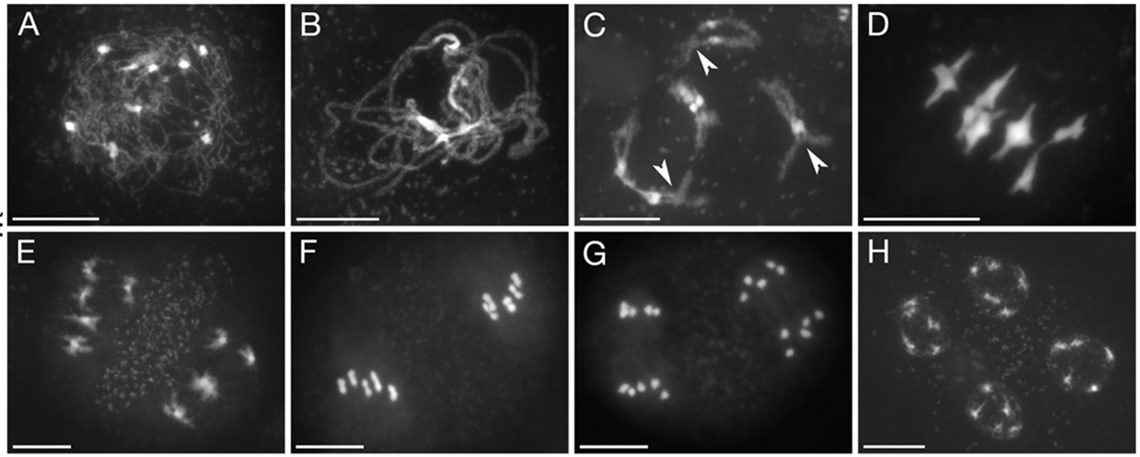
- Figure 1.3.3 Eight microscope images, labelled A to H, show the various steps of meiosis in Arabidopsis cells with a haploid number of five. Panels A-C show different stages of prophase I, each with an increasing degree of chromosome condensation. The subsequent phases are metaphase I (D), telophase I (E), metaphase II (F), anaphase II (G), and telophase II (H). [Back to Figure 1.3.3]
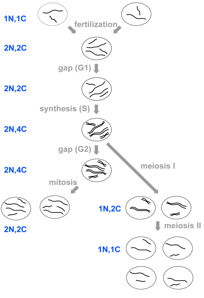
- Figure 1.3.4 A schematic diagram: DNA, represented by simple lines, outlining the changes in the chromosome number during various stages of the cell cycle, starting with gametes (haploid number), moving through fertilization (diploid number), followed by DNA replication during the S phase (synthesis phase), and showing two possibilities for cell division, with mitosis ending up with two diploid number daughter cells and mitosis ending up with four haploid number cells. [Back to Figure 1.3.4]
- Figure 1.3.5 A pair of homologous chromosomes, one coloured green and the other coloured blue, form a synaptonemal complex, and show the crossing over of non-sister chromatids to produce recombinant chromosomes post-crossover. [Back to Figure 1.3.5]
- Figure 1.3.6 Three parts: 1) a pair of homologous chromosomes, which have already been duplicated, one coloured pink and the other coloured blue; 2) The two chromosomes associate and cross over non-sister chromatids; 3) There are four resulting chromatids — two are non-recombinant and two are recombinant. [Back to Figure 1.3.6]
- Figure 1.3.7 The primary oocyte (one cell) results in the secondary oocyte (two cells) and these form polar bodies (three cells) and a mature ovum (one cell); the primary spermatocyte (one cell) forms the secondary spermatocytes (two cells), and these form the spermatids (four cells). All cells are represented by simple circles. [Back to Figure 1.3.7]

.svg)
.svg&oldid=507847250)