3.5 Appearance of a Typical Nuclear Chromosome During the Cell Cycle
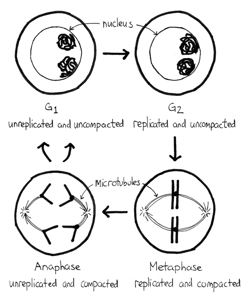
If we follow a typical chromosome in a typical human cell, it alternates between unreplicated and replicated states, and between relatively uncondensed and condensed. The replication is easy to explain. If a cell has made the commitment to divide, it first needs to replicate its DNA. This occurs during S phase. Before S phase, chromosomes consist of a single piece of double-stranded DNA and after they consist of two identical double-stranded DNA molecules.

The condensation is a more complex story because eukaryotic DNA is always wrapped around some proteins. Figure 3.5.1 shows the different levels commonly found in cells. During interphase, a chromosome exists mostly as a 30 nm fibre. This allows it to fit inside the nucleus and still have the DNA be accessible for enzymes performing RNA synthesis, DNA replication, and DNA repair. At the start of mitosis, these processes halt and the chromosome becomes even more condensed. This is necessary so that the chromosomes are compact enough to move to the opposite ends within the cell. When mitosis is complete the chromosome returns to its 30 nm fibre structure. Recall that each of our cells has a maternal and a paternal chromosome 1. Figure 3.5.2 shows what these chromosomes look like during the cell cycle.

Media Attributions
- Figure 3.5.1 Chromatin Structures by Richard Wheeler at en.wikipedia, CC BY-SA 3.0, via Wikipedia
- Figure 3.5.2 Original by M. Harrington (2017), CC BY-NC 3.0, MRU Open Genetics Lectures
Reference
Harringon, M. (2017). Figure 8. Appearance of maternal and paternal chromosome 1 …[diagram]. In Locke, J., Harrington, M., Canham, L. and Min Ku Kang (Eds.), Open Genetics Lectures, Fall 2017 (Chapter 15, p. 6). Dataverse/ BCcampus. http://solr.bccampus.ca:8001/bcc/file/7a7b00f9-fb56-4c49-81a9-cfa3ad80e6d8/1/OpenGeneticsLectures_Fall2017.pdf
Long Descriptions
- Figure 3.5.1 The various levels of chromosome compaction and architecture in a cell are depicted as follows: A DNA double helix is followed by the combination of DNA and core histones, then the beads-on-a-string structure, followed by more histone additions, to form the 30nm fiber. This is then combined with scaffolding proteins to eventually result in the typically shaped and presented chromosome. [Return to Figure 3.5.1]
- Figure 3.5.2 The appearance of chromosomes during the G1 and G2 growth stages of the cell cycle is different from chromosomes in the anaphase and metaphase of mitosis. During the growth phases, the chromosomes are unreplicated and uncompacted, compared to anaphase and metaphase, where the chromosomes are compacted and condensed, and spindle fibres are shown to be present in the cell during these stages of mitosis. [Return to Figure 3.5.2]

