3.3 Mitosis
During the S-phase of interphase the chromosomes replicate so that each chromosome has two sister chromatids attached at the centromere. After S-phase and G2, the cell enters Mitosis. The first step in mitosis is prophase, where the nucleus dissolves and the replicated chromosomes condense into the visible structures we associate with chromosomes. Next is metaphase, where the microtubules attach to the kinetochore and the chromosomes align along the middle of the dividing cell, known as the metaphase plate. The kinetochore is the region on the chromosome where the microtubules attach. It contains the centromere and proteins that help the microtubules bind. Then in anaphase, each of the sister chromatids from each chromosome gets pulled towards opposite poles of the dividing cell. Finally in telophase, identical sets of unreplicated chromosomes (single chromatids) are completely separated from each other into the two daughter cells, and the nucleus re-forms around each of the two sets of chromosomes. Following this is the partitioning of the cytoplasm (cytokinesis) to complete the process and to make two identical daughter cells. An acronym to remember the main stages of mitosis is iPMAT, where the little (lowercase) i stands for interphase.

Take a look at the following video, Cell Biology |Cell Cycle: Interphase & Mitosis, by Ninja Nerd (2018) on YouTube, which discusses the various stages of the Cell Cycle.
You should note that this is a dynamic and ongoing process, and cells don’t just jump from one stage to the next.

Keep in mind the following points that outline the importance and significance of mitosis:
- Keeps chromosome number constant
- Maintains genetic stability in daughter cells
- Helps in growth and development of the zygote
- Helps in repair and regeneration
- Restores nucleo-plasmic ratio
- Checks cell size and maintains a favourable surface area/volume ratio.
How Mitosis Helps to Maintain Genetic Stability
Mitosis results in the splitting of replicated chromosomes during cell division and facilitates the generation of two new identical daughter cells. Given that chromosomes form from parent chromosomes by making exact copies of their DNA, mitosis helps in preserving and maintaining the genetic stability of a particular population.
Watch the video, Animation How the Cell Cycle Works, by Marcin Gorzycki (2018) on YouTube, which discusses the various stages of Mitosis.
Media Attributions
- Figure 3.3.1 0331 Stages of Mitosis and Cytokinesis by Betts et al. (2016), OpenStax, CC BY 4.0, via Wikimedia Commons
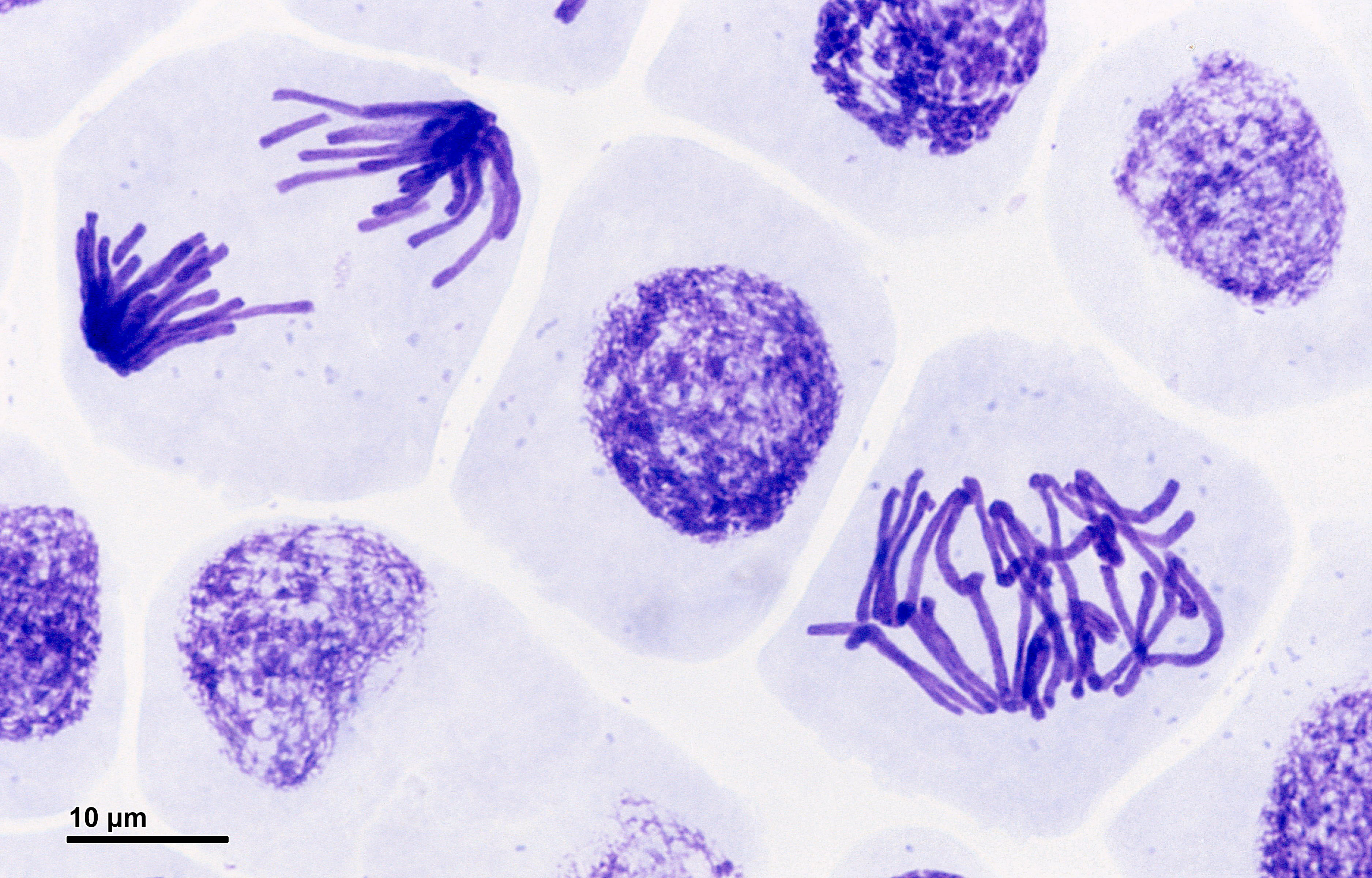
- Figure 3.3.2 Pressed root meristem… by Doc. RNDr. Josef Reischig, CSc. (2014), Author archives/ Mantis, CC BY-SA 3.0, via Wikimedia Commons
References
Betts et al. (2016, October 21). Figure 3.32 Cell division: Mitosis followed by cytokinesis [digital image]. Anatomy and Physiology. OpenStax. https://openstax.org/books/anatomy-and-physiology/pages/3-5-cell-growth-and-division
Marcin Gorzycki. (2013, March 14). Animation how the cell works (video file). YouTube. https://youtu.be/g7iAVCLZWuM”>https://youtu.be/g7iAVCLZWuM”>https://youtu.be/g7iAVCLZWuM
Ninja Nerd. (2018, January 7). Cell biology | Cell cycle: Interphase & mitosis (video file). YouTube. https://youtu.be/LUDws4JrIiI”>
Reischig, J. (2014). Mitosis: pressure; root meristem; Vicia faba, A, P, 1500x [digital micrographic image]. Mantis. https://www.mantis.cz/mikrofotografie/pages/261_10.html
Long Descriptions
- Figure 3.3.1 The various stages of mitosis are shown, which include the prophase, prometaphase, metaphase, anaphase, telophase, and cytokinesis. Please refer to the text for details of each stage, and pay special attention to the description of the movement of chromosomes and their occurrences during mitosis. [Back to Figure 3.3.1]
- Figure 3.3.2 A vivid slide presents purple-colored stained chromosomes in root cells, which all exemplify various stages of mitosis. In some cells, chromosomes are diffuse; in other cells, chromosomes are lined up at the equator; and in other cells, chromosomes are shown being pulled apart to opposite poles of the cell. [Back to Figure 3.3.2]

